
【 Japanese / English 】
Research
The goal of our research is to use neurophysiology to examine and explain the effects of rehabilitation in those with brain and spinal cord disorders. We aim to contribute to the development of these rehabilitation methods by explaining brain function and pathologies through EEG research.
In addition, we are conducting research to clarify emotion (pre-consciousness) and affection (consciousness).
Introduction to our research ⇒ click here
Technique
ERP
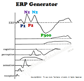
Event-related potentials (ERPs) are potentials of a few dozen microvolts measured as an electroencephalogram (EEG) component that is a result of neuronal activity in the cerebral cortex. Among various EEG components, the ERP is an intrinsic potential that reflects the subject’s cognitive response towards an event (stimulus). In ERPs, characteristic components appear at specific latencies in response to various cognitive processes, therefore it is widely used in the measurement of the processes of “sensation, perception, and cognition.”
LORETA
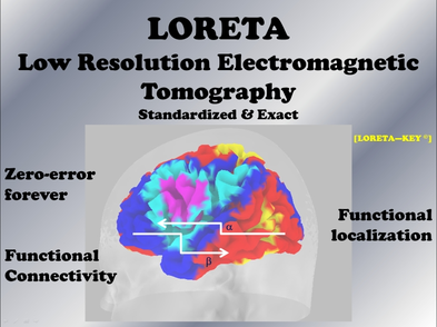
LORETA (low resolution (brain) electromagnetic tomography analysis) is a functional imaging analysis of the brain developed by R.D. Pascual-Marqui et al. LORETA allows us to superimpose intracerebral neural activity observed by EEG and magnetoencephalography onto a brain atlas (standard brain).
LORETA was first reported in 1994; however, sLORETA and eLORETA, released in more recent years, not only draw the potential distribution of brain regions divided into 6239 voxels, but also analyze functional connectivity (LORETA connectivity) and networks (eLORETA-ICA) between different brain regions. Comparison to a database of healthy individuals (built in the program) is also possible with sLORETA and eLORETA.
Microstate segmentation
(Michel MC, et al. 2009)
Microstate segmentation analysis is a method published by Lehmann et al that analyzes data obtained from EEG measurements in clusters, and determines intervals in which nearly stable topography appears continuously, or in other words, it groups them as “microstate segments” of the brain.

Projects
Neuro-reorganization
The following are some of our efforts toward neuro-reorganization.
【 Development of a neurorehabilitation system that synchronizes kinesthetic illusions and imagery 】
We have been working on the development of treatments aimed at regaining cerebral function in patients with sensorimotor disorders caused by stroke. Physical paralysis caused by stroke is characterized by declining physical awareness (corporeal representation of one’s self in the brain) that one’s body is one’s own, in addition to an increased difficulty in intentional or voluntary movements.
A “physical control system” makes the execution of movements possible in humans. When brain function is impaired, not only does the input processing of external sensory information become dysfunctional, but also the system in the brain that predicts and identifies feedback malfunctions. Furthermore, the awareness created in the body that “one is moving his/her own body” diminishes with higher-order meta-representational deficits.
For these deficits, we developed and examined the effects of imagery neurofeedback based multi-sensory systems (iNems) training aimed at improving the ability to identify movements of the limbs predictably and intentionally created in the brain, as well as actual sensorimotor information.
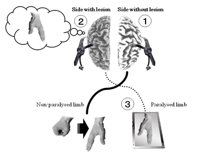
- iNems -
①The non-paralyzed limb performs the intended movement; the motor preparatory electric potential is measured and analyzed from the electrode positions in the sensorimotor-related region in the intact hemisphere in order to sense the frequency pattern.
②The patient is asked to activity imagine the same movement in the paralyzed limb, and the brain wave activity in the sensorimotor-related region in the hemisphere containing the lesion at that time is recorded.
③When the frequency pattern sensed from the intact hemisphere matches the frequency pattern from the hemisphere containing the lesion, the image on the monitor moves to provide visual feedback.
The neural activity pattern of the brain during the imaging of a movement must be captured within multiple frequency bands. We examined neural activity during movement imagination in the brain by analyzing the frequency patterns appearing within the theta, alpha, and beta wave bandwidths (Patent Application Laid-Open Publication: 2017-102504).
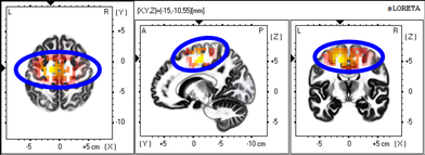
As a result of a 6-week training regimen, neural activity in sensorimotor-related areas (inside the blue circle) centered around the supplementary motor area was enhanced within the alpha bandwidth (mu waves) during imaging. In addition, the sense of agency and ownership, frequency of the use of the affected limb, and QOL also improved. Going forward, we plan to apply this training program to a larger population to further examine its effects.
・Kodama T, et al., Clin EEG Neurosci. 2019
【 Development of a neurorehabilitation system that synchronizes kinesthetic illusions and imagery 】
Our work has been done to establish treatment methods for reorganizing brain functions in patients with sensorimotor disorders caused by stroke or cervical spinal cord injury.
In many previous studies, real-time feedback has reported the use of visual, electrical and auditory stimuli as an approach to treat sensorimotor impairments of a hand. This state (in fact, there is a time delay) increases the possibility of synchronous processing of the matching between motor intention and sensory feedback information in the brain, leading to reorganization of sense of motor subjectivity and body ownership. This temporal congruence was reported to increase activity in the premotor cortex and corticospinal tracts, leading to improved upper extremity performance.
However, it is necessary for the hand to confirm the coordinates of the target object by visual information and control the friction (dynamic friction) generated within the finger abdomen with the detected object by muscle activity in order to execute skillful motions. The idea is to make precise sense of these objects and learn them in the brain with feedback stimuli reported so far.
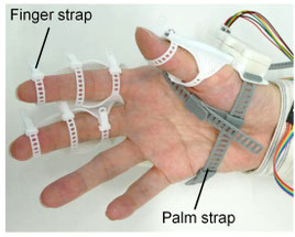
We performed effectiveness verification using Yubirecorder (developed by Tec-Gihan), a system that provides real-time compensatory sensory (vibration) feedback of friction information generated when an object is touched (conceived and developed by Professor Yoshihiro Tanaka of Nagoya Institute of Technology).
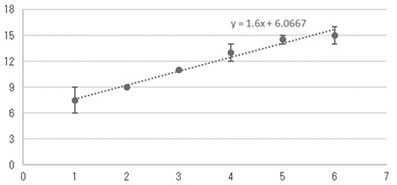
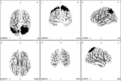
For 6 weeks of training in subjects with cervical spinal cord injury, an improvement was observed in peg-test evaluation (left figure), and comparison of EEG activity before and after intervention (right figure) showed improved neural activity in parietal association cortex (black region). Further improvement was also observed in the sense of motor independence, frequency of use of the influenced limb, and quality of life. Similar results were obtained for stroke patients.
We plan to conduct this training for more people in the future and validate the effectiveness of this training in depth.
・Kitai, Kodama et al., Brain Sci. 2025
・Kitai, Kodama et al., Brain Sci. 2021
・Kodama & Kitai, IntechOpen. 2023
・Kitai, Kodama et al., Brain Sci. 2021
Neuro-mechanics
The following are some of our efforts toward neuro-mechanics.
【 Investigating the Neural Mechanisms of Self-Controlled and Externally Controlled Movement with a Flexible Exoskeleton Using EEG Source Localization 】
In neurorehabilitation for sensorimotor dysfunctions, combining motor imagery with self-controlled active movement is crucial; however, performing voluntary movements is often difficult for patients with severe motor impairments.
To address this, we developed "flexEXO," a soft and flexible exoskeleton device designed to assist finger movements, and investigated cortical activation using EEG during self-controlled (SCC) and externally controlled (OCC) grasping tasks.

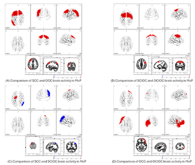
The results revealed that self-controlled tasks, where participants initiated the device's movement, showed higher event-related desynchronization (ERD) and robustly engaged motor planning and execution regions such as the primary motor cortex (M1) and supplementary motor area (SMA).
In contrast, externally controlled movements enhanced activation in the inferior parietal lobule (IPL) and secondary somatosensory cortex (S2), which are associated with sensory feedback processing.
These findings demonstrate that self-controlled motor training facilitated by flexEXO effectively engages essential motor networks, highlighting its potential to promote neural plasticity and motor learning in rehabilitation.
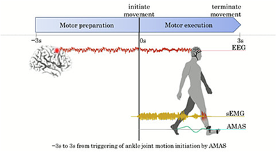
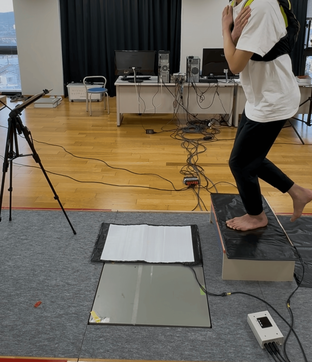
【 The Link Between Motor "Preparation" and "Execution" Determines Walking Ability: A Novel EEG-Based Gait Assessment 】
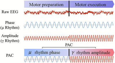
Ankle motor control is essential for the recovery of gait after a stroke. However, accurately assessing this ability in patients with severe paralysis has been challenging. We focused not only on the actual movement but also on the neural network connection between motor "preparation" (intention/planning) and "execution" (Phase-Amplitude Coupling: PAC). In this study, we simultaneously measured electroencephalography (EEG) and electromyography (EMG) during active ankle dorsiflexion.
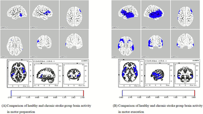
The figure on the left shows the sLORETA images, illustrating the differences in brain activation areas (e.g., insula, middle temporal gyrus) between healthy controls and stroke patients during motor preparation and execution.
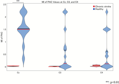
The figure on the right is the violin plot showing the stark contrast in PAC-MI values between the two groups, providing a clear visual representation of the findings.
We found that while healthy individuals exhibit a strong coupling between motor preparation and execution (high PAC-MI values), this connection is significantly diminished in stroke patients. This brain "preparation-execution coupling" is strongly associated with actual walking speed and gait performance. It holds great promise as a novel neurophysiological biomarker for early rehabilitation assessment and future neurofeedback interventions for gait reconstruction.
【 The Impact of Verbal Encouragement on Neural Networks in Children with Cerebral Palsy: The Science of Communication in Rehabilitation 】
In pediatric rehabilitation, eliciting patient motivation is essential for improving motor and cognitive functions.
However, objectively evaluating how "verbal encouragement" affects the internal state of children with severe motor and intellectual disabilities due to cerebral palsy (CP-SMID), who often face communication challenges, has been a long-standing issue. We measured the electroencephalogram (EEG) of these children while their "mothers" and "physical therapists (PTs)" provided verbal prompts using different tones (e.g., informing vs. asking).
We then visualized the responses of their brain networks using eLORETA-ICA frequency analysis.
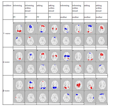

The results revealed that the activated brain frequency bands (theta, alpha, beta) and regions differed significantly depending on who was speaking and the tone used. For instance, maternal encouragement tended to facilitate internal information processing and behavioral engagement (fostering a sense of security and self-awareness). In contrast, encouragement from the PT appeared to promote emotional regulation and the cognitive processing required to grasp the meaning and intention of the words.
This study provides neuroscientific evidence of how everyday verbal interactions impact a child's emotions and motivation, directly contributing to the development of more effective, personalized rehabilitation approaches.
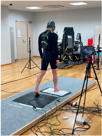
【 Elucidating Motor Learning Mechanisms Based on Predictive Control and Self-Reflection in Jump-Landing Movements 】
"Jump-landing movements," frequently performed in sports like volleyball and basketball, are highly associated with sports injuries such as anterior cruciate ligament (ACL) tears and ankle sprains. To prevent injuries and facilitate an early return to sports, "motor learning"—the rapid acquisition of safe landing mechanics—is essential.
In this study, we went beyond traditional biomechanical evaluations and utilized electroencephalography (EEG) to investigate the central nervous system mechanisms underlying motor learning.
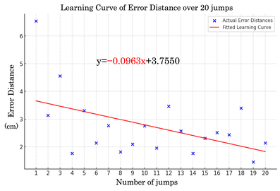
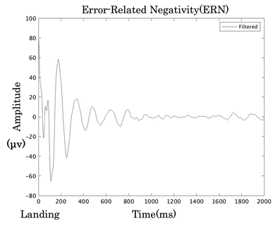
Specifically, we focused on three neural markers: the Bereitschaftspotential (BP), reflecting pre-movement "predictive control"; the posterior parietal cortex (PPC), involved in sensory integration; and the error-related negativity (ERN), reflecting post-movement "self-reflection" (error detection). Our analysis revealed that individuals with higher pre-movement BP and PPC activity corrected landing errors more quickly, indicating faster motor learning.
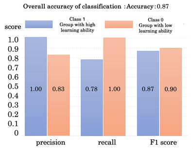
Furthermore, by employing machine learning (Support Vector Machine: SVM), we successfully predicted individuals' high or low motor learning abilities with high accuracy (87%) based on their ERN amplitudes.
These findings provide crucial insights for enhancing athletic performance and developing novel assessment and training methods in sports rehabilitation.
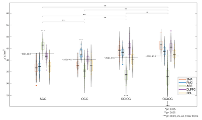
【 Elucidating the Neural Network and Error Monitoring Mechanisms Underlying Step Accuracy 】
The ability to accurately place the foot during the initial step of gait initiation is crucial for maintaining postural stability and preventing falls. However, the detailed neural mechanisms governing this instantaneous movement have remained largely unclear.
By combining mobile electroencephalography (EEG) with high-spatial-resolution eLORETA-ICA and microstate segmentation analysis, we have successfully captured real-time brain activity networks during stepping movements.
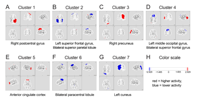
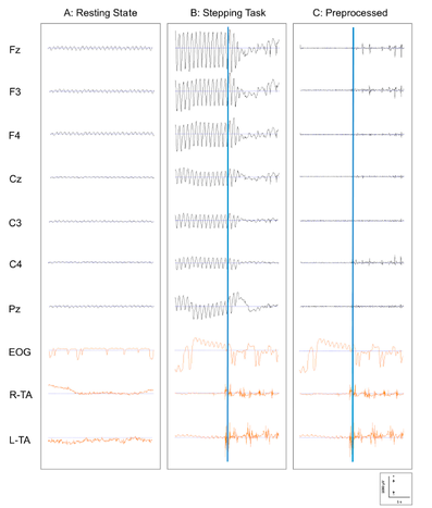
Our research revealed that individuals with high stepping accuracy exhibit specifically amplified theta-band activity in the anterior cingulate cortex (ACC). This indicates a robust "error monitoring function" wherein the brain instantaneously detects and corrects deviations in foot placement. Furthermore, we proposed the existence of a "distributed neural network" involving the suppression of mu and beta waves in the sensorimotor areas (facilitating smooth motor execution) and enhanced visuospatial processing in the parieto-occipital regions.
These findings contribute to the creation of novel biomarkers for objectively assessing fall risk in older adults and patients with neurological disorders, as well as the development of targeted rehabilitation strategies.
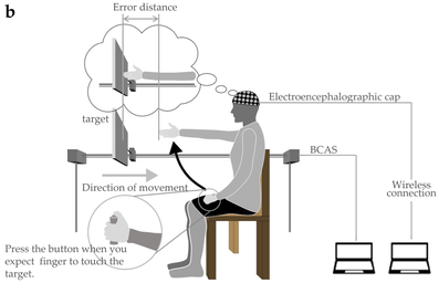
【 Capturing Real-Time Changes in Body Awareness: Development and Validation of the Novel Body Cognition Assessment System (BCAS) 】
In sensorimotor dysfunction following neurological disorders such as stroke, "body cognition"—which encompasses the sense of body ownership and the sense of agency—is often altered, significantly hindering activities of daily living (ADLs).
However, conventional assessment methods are largely static or rely on indirect questionnaires, making it difficult to directly capture the "latent body cognition" that dynamically changes during actual movement.
To address this, we developed a novel Body Cognition Assessment System (BCAS), in which participants press a button to stop an approaching target at the exact moment they perceive it has reached their arm's length (reach distance).
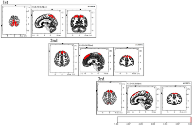
When tested on healthy participants, the error distance (BCAS value) fluctuated across repeated trials. Simultaneous electroencephalogram (EEG) analysis revealed that the activated brain regions shifted dynamically across trials: from the superior parietal lobule (associated with a sense of body ownership) in the first session, to the dorsolateral prefrontal cortex (sense of agency) in the second, and to the supplementary motor area (body-motor imagery) in the third.
This suggests that the BCAS successfully and sensitively captures the immediate updating of body cognition during the early stages of motor learning. This system holds great promise as a new objective assessment tool in rehabilitation, contributing to the functional recovery of patients with neurological disorders.
【 Examination of neural activity in the brain during kinesthetic illusions caused by vibratory stimulation 】
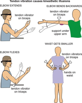
* For instance, when vibratory stimulation is applied to a muscle or tendon that flexes the elbow, a sensation or illusion that the elbow is extended is generated (kinesthetic illusion). (Janet, 2013)
We have analyzed the effects of kinesthetic illusions (above figure) generated by vibratory stimulation on brain function using LORETA and using the mu rhythm (aka mu waves), which is are brainwaves that are said to be attenuated during both actual exercise and exercise image recall, as a neurophysiological indicator.
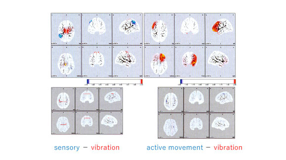
The results showed that mu waves were attenuated (ERD) in the sensorimotor region (including the supplementary motor area) of the cerebral cortex during vibratory stimulation and exercise. Furthermore, a comparison of the two conditions indicated that there was no significant difference in the neural activity in the region. This suggests that the quantitative neural activity in the region is similar during both kinesthetic illusions and exercise. Moreover, it indicates that mu waves are attenuated in patients with impaired brain function (i.e. cerebrovascular disease) as well as in healthy patients.
On the other hand, it is thought that kinesthetic imaging ability is important for the generation of kinesthetic illusions in the brain. We believe that detailed verification of these associations and further examination of the effects will contribute to the creation of future neurorehabilitation protocols.
【 The effects of environmental colors on cognitive function 】
We have compared and examined the effects of environmental colors such as red and green on visual tasks relating to cognitive function by analyses using the P300 component, an event-related potential, and LORETA. The results showed that red increased the maximum amplitude of the P300 component and reduced latency.
In addition, LORETA showed significantly higher neural activity in the inferior temporal gyrus, amygdaloid nucleus, anterior cingulate gyrus, and Brodmann area 46, which are considered to be strongly related to emotional and executive functions.
Through these examinations, it was shown that cognitive function is influenced by the color in the patient’s environment.
・Kodama et al. Brain science and mental disorders. 2007
・Kodama et al., J Neurotrauma. 2010
・Kodama et al., JPTS. 2016
Neuro-feedback
The following are some of our efforts toward neuro-feedback.
【 Examination of the effects of neurofeedback training in patients with intractable pain 】
<What's neuro-feedback trainig ?>
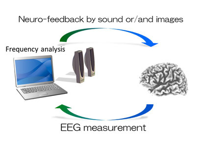
In neurofeedback training, brain function is measured and analyzed using brainwaves (as signals obtained from a living body), and the results are synchronously returned to the patient, thereby attempting to create a stable and improved neurological state of by himself/herself. Neurofeedback training is used for individuals suffering from chronic pain or anxiety symptoms.
In this training, brainwaves are monitored and frequency analysis is performed to understand the state of the brain. During the resulting feedback, auditory or visual signals are given when brain activity is in a positive state (i.e. when the alpha waves that appear when one is relaxed become dominant). This lets the trainee know that the state of brain function at that time is positive.
By repeating this exercise, the trainee naturally learns by experience (conditioning), and he/she becomes able to constantly maintain a positive state of the brain outside the training settings.
The figure below is an EEG image of a patient with complaints of chronic pain and anxiety.
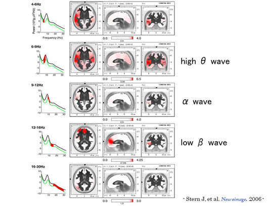
This figure shows that the anterior cingulate gyrus, dorsolateral prefrontal cortex, inferior parietal lobe, and islet regions of the brain in these patients showed higher neural activity than in healthy individuals, in particular the high theta bandwidth and the bandwidth known as beta waves.
These results indicate that chronic pain and anxiety symptoms are associated with these areas (“painmatrix”, “neuromatrix”) in recent years. Furthermore, we believe these results elucidate the brain function characteristics of this neural activity by each frequency pattern.
We have performed and examined the effects of interventional neurofeedback training on patients with intractable pain. We noted that the resting beta bandwidth diminished, and anxiety and pain were relieved.
TK-Lab.
Takayuki Kodama, Department of Physical Therapy, Faculty of Health Sciences, Kyoto Tachibana University,
34 Yamada-cho, Oyake, Yamashina-ku, Kyoto 607-8175 Japan.
Copyright©2013 TK-Lab, Kyoto Tachibana University All Rights Reserved.